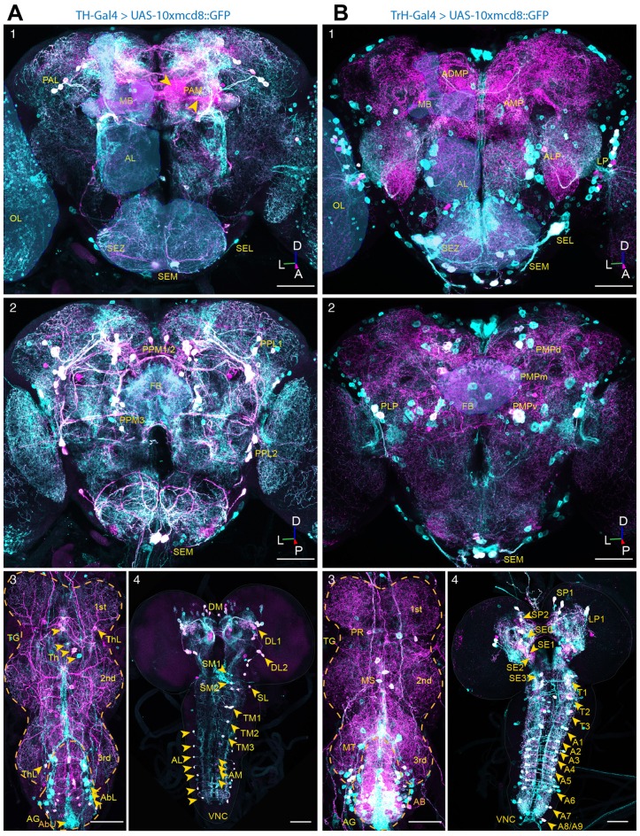
Figure 1.
Dopaminergic and serotoninergic neurons of the adult Drosophila central brain. (A) In situ co-immunostainings with anti-GFP (green) and anti-tyrosine hydroxylase (TH; magenta) antibodies in whole-mount nervous tissues of TH-Gal4 >10xUAS-mCD8<GFP flies. Co-localizations merge both colors in white, showing driver-targeted somata of the subesophageal medial (SEM), lateral (SEL), the protocerebral medial (PAM) and lateral (PAL) clusters and projections in anterior (1) and the posterior lateral (PPL1, PPL2) and medial clusters (PPM1/2, PPM3) in the posterior (2) brain, as well as the lateral (ThL) and medial cell cluster (Th) in the thoracic ganglion (TG) and the lateral (AbL) and medial (AbU) situated clusters in the abdominal ganglion (AG) (3) of adult wild-type Drosophila. In the central nervous system (CNS) of LIII-Larvae (4) the dopaminergic system consists of one dorso-medially (DM), two dorso-laterally (DL1, DL2) within the hemispheres, three clusters in the medial (SM0, SM1, SM2) and one cluster in lateral (SL) subesophageal zone (SEZ), three medially situated cluster in the thoracic (TM1, TM2, TM3) clusters and an array of neurons in the lateral (AL) and medial (AM) part of the abdominal part of the ventral nerve cord (VNC). (B) In situ co-immunostainings with anti-GFP (green) and anti-5-HT (magenta) antibodies in whole-mount nervous tissues of TrH-Gal4>10xUAS-mCD8<GFP flies, showing the clusters in the lateral lateral protocerebrum (LP), the anterior medial protocerebrum (AMP), anterior lateral protocerebrum (ALP) and the anterior dorso-medial protocerebrum (ADMP) and the medially (SEM) and laterally (SEL) situated clusters in the subesophagealsubesophageal zone (SEZ) in the anterior (1) and the clusters situated in the dorsal (PMPd), medial (PMPm) and ventral posterior protocerebrum as well as the dorsally situated clusters in the medial (SEM) and lateral (SEL) subesophageal ganglion (2) and the clusters in the por-, (PR), meso- (MS) and meta- (MT) thoracic neuromere and the AG (AB) (3) of an adult fly. The CNS of a LIII-Larva (4) of three clusters in the supresophageal ganglion (SP0, SP1, SP2), one in the LP1 and four cell clusters in the SEZ (SE0, SE1, SE2, SE3). The VNC contains three clusters of 5-HT producing neurons in the thoracic (T1, T2, T3) and an array of nine symmetrically organized clusters in the abdominal neuromere (A1–A9). Overlay in white correspond to driver-targeted serotoninergic cell bodies (MB, mushroom body; FB, fan-shaped body; OL, optic lobe; SEZ, subesophageal zone; AL, antennal lobe; TG, thoracic ganglion; AG, abdominal ganglion; D, dorsal; L, lateral; P, posterior; A, anterior). Scale bars: 50 μm.

