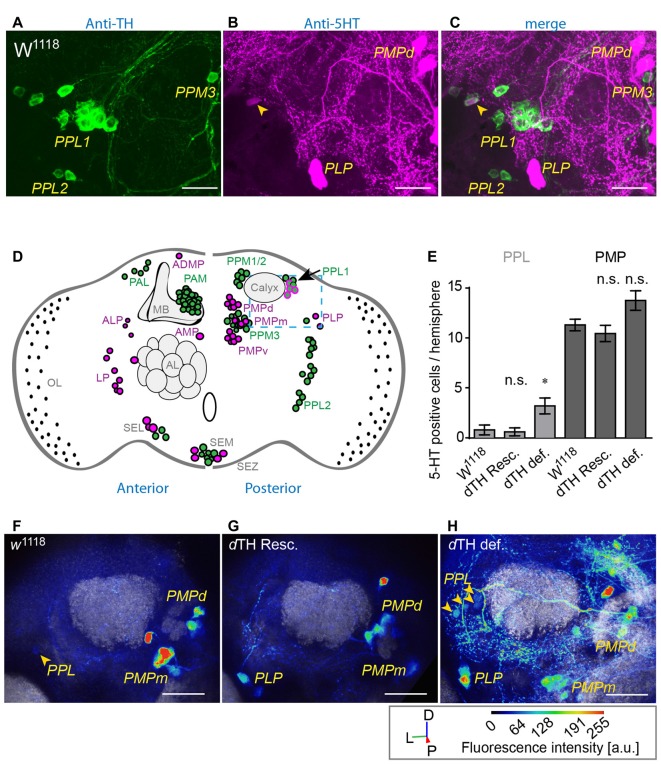
Figure 3.
DA-deficient flies show increased 5-HT immunoreactivity (IR) in TH-producing neurons of the posterior lateral protocerebrum (PLP). (A) TH- (green) and (B) 5-HT- immune reactive neurons (green) in the posterior lateral protocerebrum (PPL) and the posterior medial protocerebrum (PMP) in w1118. Merge is shown in (C). Scale bars: 10 μm. W1118 control flies show a small number of faintly 5-HT-immunoreactive neurons (magenta) among (A) DA-producing PPL1 neurons (C, arrow heads). (D) Schematic overview of the TH (green) and 5-HT (magenta) immune reactive neurons in the adult Drosophila brain. In the PPL (blue dashed line) w1118 control flies show a small number of 5-HT-immunoreactive neurons (magenta) among DA-producing neurons (green) in the PPL1 cluster (arrow). (E) DA-deficient flies show increased numbers of 5-HT-producing neurons in the PPL when compared to w1118 controls and dTH-rescue flies (Dunn’s multiple comparison test against w1118, n > 5). (F) 5-HT immune reactive neurons (green) in the PPL and the PMP in w1118, genomic dTH rescue (G, dTH Resc.) and DA-deficient flies (H, dTH def., arrow head) (D, dorsal; L, lateral; P, posterior) Scale bars: 20 μm. n.s.: p > 0.05; *p < 0.05.

