Figure 6.
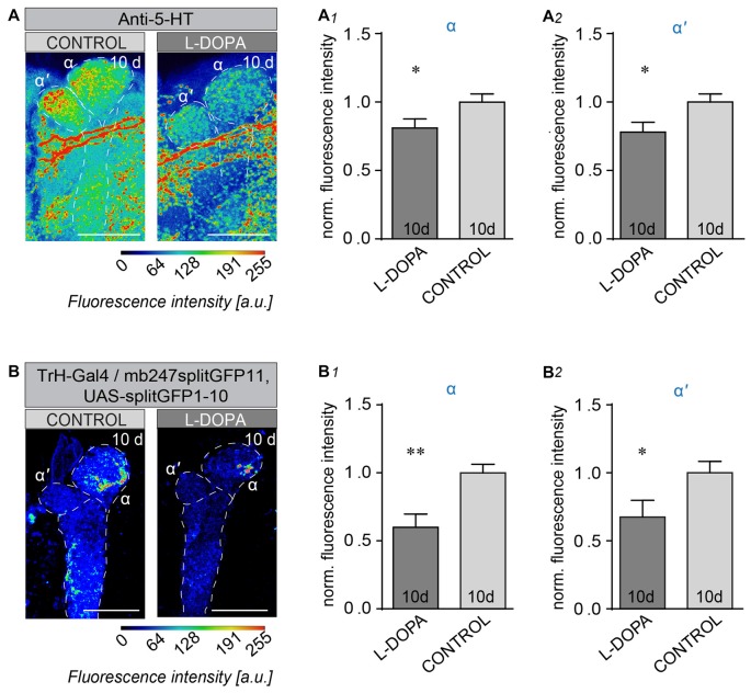
Long-term L-DOPA treatment alters 5-HT neuron innervation to their MB target regions. (A) Innervation patterns of 5-HT neurons to the α/α’-lobe of the MB. Projection of 5-HT producing neurons onto α/α’ lobes of the MB under control conditions and after 10 days L-DOPA administration. Fluorescence intensity analysis of the 5-HT IR in α-lobes (A1) and α’-lobes (A2; unpaired students T-test, n < 15). (B) Reconstituted splitGFP between TrH-positive 5-HT producing neurons and the MB vertical lobes in wild-type flies under control conditions (left) and after 10-day L-DOPA treatment (right). Fluorescence intensities are indicated by false colors. Fluorescence intensity analysis of the reconstituted splitGFP signal in the tips of α (B1) and α’ lobes (B2). 5-HT innervations are strongly reduced by L-DOPA treatment compared to non-treated control flies (Unpaired students T-test, n < 11). Scale bars: 20 μm. n.s.: p > 0.05; *p < 0.05; **p < 0.01.

