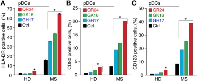
Figure 4.

Expression of the cell surface markers by plasmacytoid dendritic cells (pDCs) exposed to the individual myelin basic protein (MBP) peptides comprising the liposomal peptide therapeutic. The pDCs from the healthy donors (HD) and multiple sclerosis (MS) patients were incubated with 5 µg mL−1 of MBP peptides GH17 (blue bars), GK16 (green bars), QR24 (red bars), or PBS (black bars) for 24 h. Percentages of human leukocyte antigen (HLA-DR)+ (A), CD80+ (B), and CD123+ (C) cells were further determined by flow cytometry. Data are presented as a percentage of positive cells within the CD45+ PBMC population. Asterisk denotes a statistically significant difference with the control. NS, non-significant.
