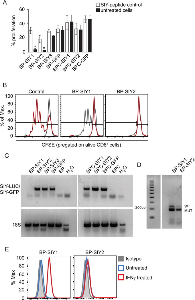
Figure 5. Immune surveillance results in a loss of immunogenicity in β-catenin-negative tumor cells but not in β-catenin-positive tumor cells.

(A) The immune-stimulatory capacity (proliferation) of tumor cell lines isolated from MC57-SIY immunized BP-SIY and BPC-SIY tumor bearing mice. Immune stimulatory potential was assessed by measuring T cell proliferation in a co-culture assay of CFSE-labeled 2C TCR transgenic T cells and untreated or SIY-peptide pulsed tumor cells. We used tumor cells isolated from antigen negative mice transduced with GFP-SIY constructs (here shown as BP-/BPC-GFP) as positive control (data are given as mean with SEM and significance was assumed with p ≤ 0.05, with * ≤ 0.05, n = 8, pooled from two independent experiments). (B) Representative examples of histograms depicting CFSE dilution for control (gray unstimulated, red transduced BP cell lines) and the two cell lines shown defective T cell stimulation. (C) RT-PCR analysis assessing the SIY-mRNA levels in the cell lines used in (A) (representative for two independent experiments). (D) Genotyping PCR confirming the presence of the SIY-LUC transgene in the BP-SIY1 cell line (WT band 235 base pairs, SIY-mutation 190 base pairs). (E) Representative histogram assessing the expression profile of MHC-I on the surface of untreated (blue) or IFN-γ-treated (red) BP-SIY1 and BP-SIY2 cell lines.
