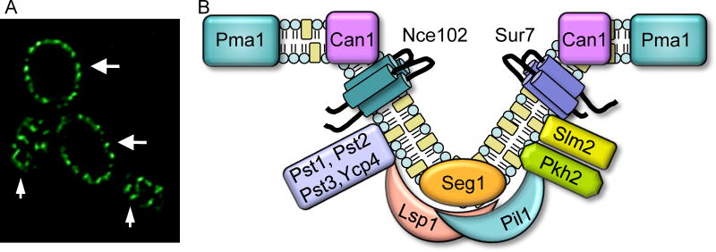
Figure 1. MCC/eisosome domains in the plasma membrane of C. albicans.
(A) MCC/eisosome domains visualized by fluorescence microscopy of C. albicans cells producing an Lsp1-GFP fusion protein. Small arrows point to regions at the top of cells where MCC/eisosome domains can be visualized as punctate patches in the plasma membrane. Larger arrows point to regions where the mid-section of the cell is in focus, in which MCC/eisosomes appear as a series of patches around the perimeter of the cell.
(B) Model for MCC/eisosome structure. Note that only a representative set of the >30 proteins that localize to MCC/eisosomes is shown.

