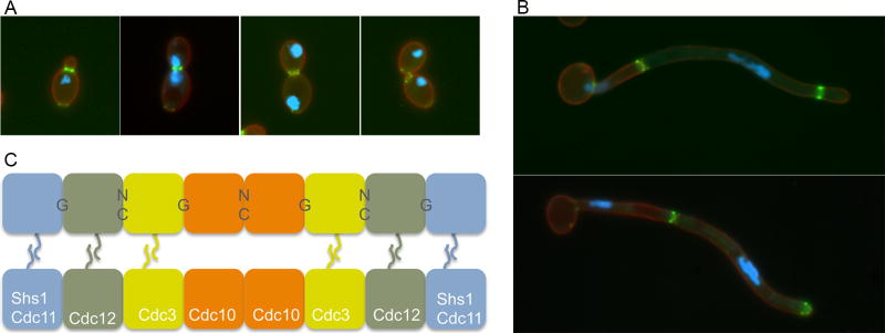
Figure 2. Septin localization in C. albicans.
(A) and (B) show septin structures in green that were visualized by fluorescence microscopy of cells that produce a fusion between the Cdc10 septin and GFP. The blue color indicates the position of the nucleus due to staining with Hoechst dye. The red color corresponds to the plasma membrane that was detected by a Pleckstrin Homology (PH) domain fused to RFP.
(A) Septin ring forms at the bud neck, converts to an hourglass, and then splits into a double ring prior to cytokinesis, and then disassembles after cell septation.
(B) Septin localization in C. albicans hyphal cells. Note that the older septin rings remain stable for a longer period of time in hyphae following cell division in the growing hyphae.
(C) Octomer model for septin filament organization in S. cerevisiae. Note that either Cdc11 or Shs1 (Sep7) can occupy the terminal positions

