
Figure 2. Epigen-bound sEGFR is monomeric.
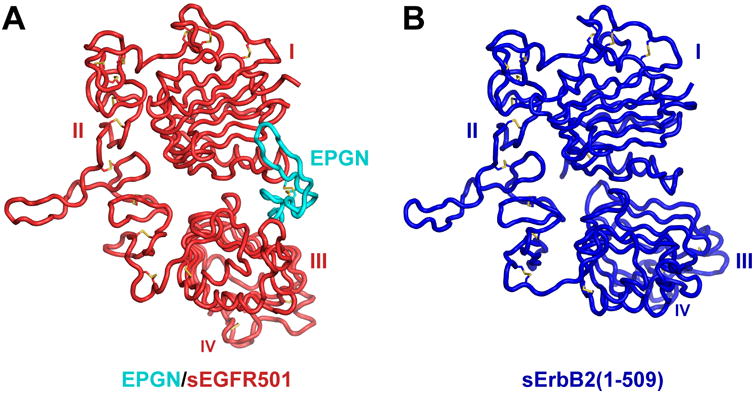
(A) Ribbon structure of EPGN-bound sEGFR501, with sEGFR501 colored red and EPGN cyan.
(B) Structure of sErbB2 (residues 1-509 – analogous to sEGFR501) in the same orientation asin (A), from PDB ID 2A91. See also Figures S2 and S3, and Table S1.