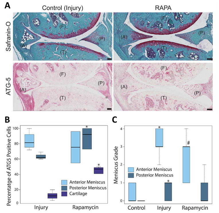
Fig. 5.
Effect of rapamycin treatment on meniscus injury and degeneration. C57BL/6J transgenic mice with surgically induced meniscus injuries and received daily intraperitoneal injections of rapamycin for 10 weeks. The knee joints were analyzed by Safranin-O staining and immunohistochemistry for ATG-5. (A) Representative images of the control (injury) and rapamycin (RAPA) treated mice knee joints stained with Safranin-O and ATG-5, showing the anterior (A) and posterior meniscus, femur (F) and tibia (T). Original magnification = 10X, Scale bar = 100 μm. (B) Quantitative analysis of ATG-5 positive cell density per 100-μm2 area. Results show significant increase in ATG-5 expressing cell number in the posterior menisci and articular cartilage. (C) Meniscus degeneration increases with injury. Rapamycin treatment does not improve degeneration in posterior meniscus. Box and whisker plots (5 mice per group); * = P < 0.05 versus control; # = P = 0.07 versus injury.

