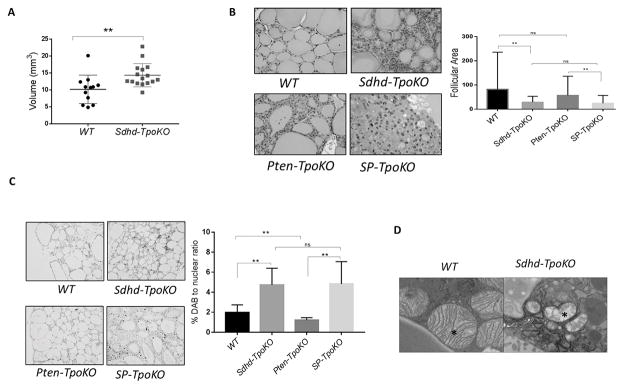
Figure 1. Sdhd deletion leads to mitochondrial defects and enhanced cellularity in vivo.
A. Thyroid volumes of WT (n=12) and Sdhd-TpoKO (n=16) mice analyzed by 3D ultrasonography at 6 months age. B. Representative images of follicles in WT, Sdhd-TpoKO, Pten-TpoKO and SP-TpoKO mice (200x magnification) at 1 year age. Quantification of the average follicular size of WT thyroids compared to Sdhd-TpoKO , and Pten-TpoKO thyroids compared to SP-TpoKO is on right. C. Ki-67 staining by immunohistochemistry in formalin fixed thyroid sections of WT, Sdhd-TpoKO , Pten-TpoKO and SP-TpoKO mice (100x magnification). Quantification of Ki-67 positive nuclei is on right. D. Mitochondrial ultrastructure (17,000x magnification) of WT and Sdhd-TpoKO mice (n=3 each) at 6 months age. Representative TEM images are shown and mitochondria are indicated by the asterisk. Error bars represents standard deviation (SD). Statistical analyses were performed by two-tailed Student’s t-test (*P value ≤ 0.05, **P value ≤ 0.01).

