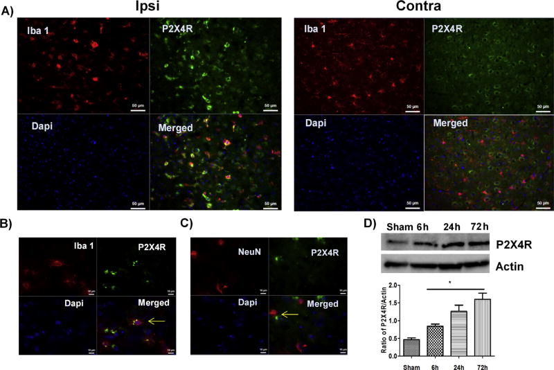
Fig. 1.
Immunolabeling of P2X4R in male WT mice three days after stroke induction. (A) P2X4R expression (green) was increased and Iba 1+ cells (red) showed an activated phenotype (round cell bodies with low processes) in the ipsilateral (stroke) versus contralateral (non-stroke) hemisphere (20x; scale bar 50 µm). (B) Ipsilateral staining: P2X4R (green) co-localized with Iba+ cells (red) (63x; scale bar 10 µm). (C) Ipsilateral staining: Co-localization of P2X4R (green) with NeuN+ve neurons (red) and DAPI (blue; marking nuclei) showed qualitatively reduced expression of P2X4R in neurons (n = 3 M WT) (63x; scale bar 10 µm) (D). Stroke led to a significant time-dependent increase in P2X4R expression in whole cell lysate from the ipsilateral brain (*p < 0.05; stroke vs. sham, one-way-ANOVA; graphs show mean + S.E.M.; n = 12; 3/group/time point; no exclusion). (For interpretation of the references to colour in this figure legend, the reader is referred to the web version of this article.)

