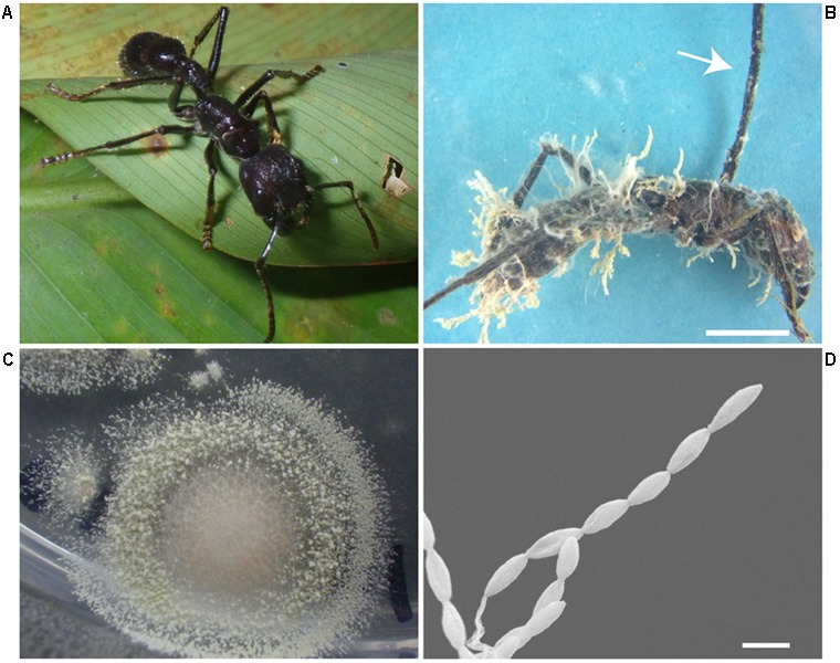
FIGURE 7.

Paraponera clavata ant parasitized by fungi. (A) Healthy P. clavata ant. (B) Infected ant, arrow pointing at the stroma that grew on the back of the ant’s head with no perithecia, note the dense yellowish aerial hyphae with chains of conidia growing on the ant’s body. Scale, 5 mm (C) Isolate BAI obtained from the P. clavata cadaver. (D) SEM micrograph, showing the conidia chains growing directly from the ant’s infected body. Scale, 2 μm.
