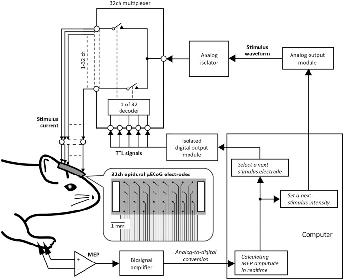
Figure 1.
Automatic motor mapping system. Stimulus current was applied epidurally through one of the 32 micro-electrocorticographic (μECoG) electrodes placed on the rat motor cortex. In the picture of the 32ch ECoG electrode sheet, black dots represent stimulus electrodes and white boxes represent stimulus grounds. Individual electrodes for cortical stimulation were selected by a 32ch multiplexer that was controlled using transistor-transistor logic (TTL) signals sent from the isolated digital output module. The stimulus waveform (e.g., duration, polarity, and intensity) was regulated by a signal sent from the analog output module. Motor evoked potential (MEP) was then stored on a computer. Thereafter, the implemented algorithm selected the μECoG electrode and the intensity value of the next stimulation, depending on the previous MEP amplitude.

