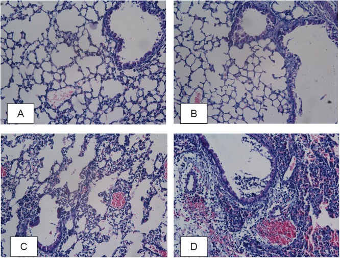
FIGURE 4.

Lung histological examination after infection. Sections of lungs stained with hematoxylin-eosin at 24 h post infection are shown (magnifications, × 400). (A) PBS group; (B) AI-2 group; (C) P. aeruginosa PAO1 group; (D) P. aeruginosa PAO1+AI-2 group.
