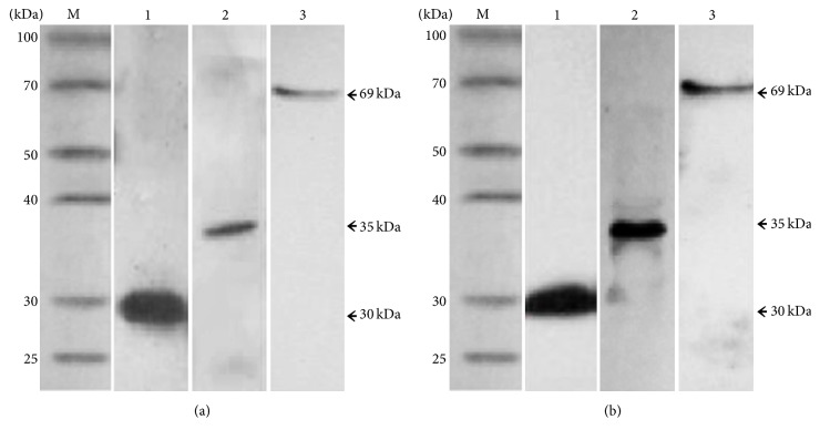
Figure 2.
Western blotting analysis of the VP1 protein and the VP1-gp120 and VP1-E2 fusion proteins. (a) Western blotting analysis using the mouse anti-His tag monoclonal antibody as the primary antibody. Lanes 1, 2, and 3 correspond to the VP1 protein and the VP1-gp120 and VP1-E2 fusion proteins, respectively. (b) Western blotting analysis using the anti-type A FMDV VP1 monoclonal antibody as the primary antibody. Lanes 1, 2, and 3 correspond to the VP1 protein and the VP1-gp120 and VP1-E2 fusion proteins, respectively.

