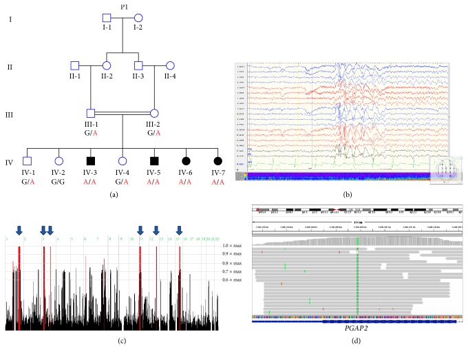
Figure 1.
Pedigree of studied kindred, EEG recordings, homozygosity mapping, and the PGAP2 variant: (a) pedigree of consanguineous Bedouin kindred studied. Below each individual are the alleles of the PGAP2 mutation, whereas G (in black) represents the wild-type allele while A (in red) represents the mutant allele. (b) EEG recordings of patient IV-5 during wakefulness showing generalized interictal spike and slow epileptiform discharges more prominent anteriorly. (c) Homozygosity-Mapper plot; blue arrows present homozygous loci shared by three affected individuals. (d) Integrative Genomic Viewer (IGV) showing the PGAP2 variant.

