Figure 4.
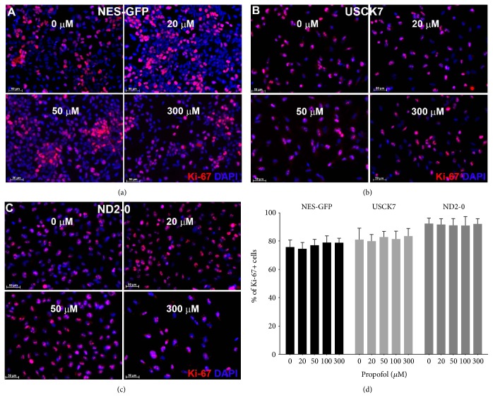
Propofol treatment for 6 h did not affect NPC proliferation. NPCs derived from three hiPSC lines were treated with propofol at different concentrations (0, 20, 50, 100, and 300 μM). Cell proliferation was assessed by Ki-67 (red) immunocytochemistry staining. Nuclei were revealed by DAPI (blue) (a, b, c). At least 1000 cells were counted for each experiment. Data were expressed as percentage of Ki-67+ cells (mean ± SD). n = 3 Ki-67 staining per treatment condition. Bar, 50 μm.

