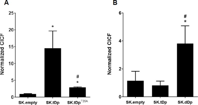
Fig. 5.

t-Darpp and Darpp-32 interact with RI. Using the proximity ligation assay, the association of t-Darpp with RIα/β in SK.empty, SK.tDp, SK.tDpT39A, and SK.dDp cells was visualized, cellular fluorescence was quantified using ImageJ, and CICF was calculated. (A) #2306 antibody that detects t-Darpp + Darpp-32 and #610165 antibody that detects RIα/β. (B) #2302 antibody that only detects Darpp-32 and #610165 antibody that detects RIα/β. Shown are the average normalized CICF values (±S.D.) from three independent wells in a single experiment. Statistical significances were evaluated using ANOVA corrected for multiple comparisons using Tukey’s post-hoc test. *p ≤0.05 compared to SK.empty; #p ≤0.05 compared to SK.tDp. Each cell line was analyzed in 1–2 independent experiments for each antibody combination.
