
Fig. 6.
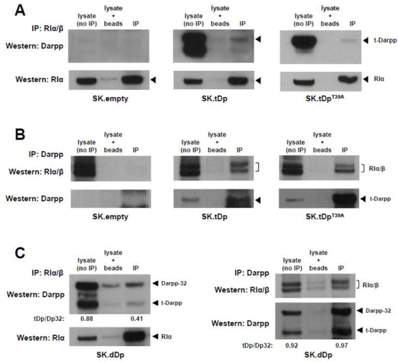
t-Darpp and Darpp-32 co-immunoprecipitate with RI. (A) Lysates from SK.empty, SK.tDp and SK.tDpT39A cells were subjected to precipitation with mouse anti-RIα/β antibody (BD Biosciences #610165) followed by Western analysis using rabbit anti-Darpp antibody (#2306, Cell Signaling). Western analysis of lysates (without prior immunoprecipitation), lysates incubated with protein G-agarose beads (no primary antibody), and immunoprecipitated lysates (IP) are shown. Stripping and re-probing for RIα (Cell Signaling antibody #5675) is shown in the bottom panels. (B) Lysates were subjected to precipitation with a rabbit anti-Darpp antibody (#2306) followed by Western analysis using mouse anti-RIα/β antibody (#610165). Stripping and re-probing for t-Darpp (#2306) is shown in the bottom panels. (C) Left, lysates from SK.dDp cells were subjected to precipitation with mouse anti-RIα/β antibody (#610165) followed by Western analysis using rabbit anti-Darpp antibody (#2306), with re-probing for RIα (#5675) in the bottom panel. Right, lysates were subjected to precipitation with rabbit anti-Darpp antibody (#2306) followed by Western analysis using mouse anti-RIα/β antibody (#610165), with re-probing for t-Darpp and Darpp-32 (#2306) in the bottom panel. The ratios of t-Darpp to Darpp-32 in lysate and IP elution lanes were quantified by measuring the bands densities using ImageJ software.