
Figure 4. EB-affected skin is infiltrated with T lymphocytes and myeloid cells.
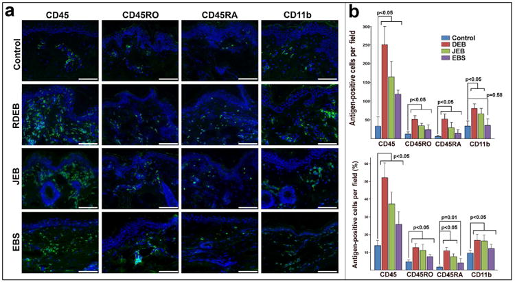
(a) Immuno-phenotyping of infiltrating leukocytes in normal (control) and non-blistering EB-affected skin using CD45, CD45RO, CD45RA and CD11b antibodies (green). EB types appear to the left of the micrographs. Detected antigens appear on the top. Blue - DAPI nuclei staining. Scale bar - 100 μm. (b) Quantitation of skin-infiltrating CD45RO+ and CD45RA+ T cells and CD11b+ myeloid cells. Analysis was done on 5 random sections from 3 independent biopsies of control and EB-affected skin. Data are presented as number and percentage of antigen-positive cells per microscopic field ± SD. Infiltration was compared using t-test. p-value ≤ 0.05 was considered statistically significant.