
Figure 5. EB-affected skin is infiltrated with CXCR2+ leukocytes.
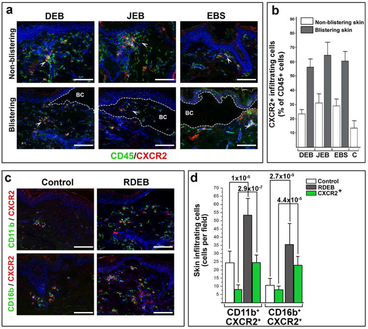
Infiltration of the DEB, JEB and EBS-affected blistering and non-blistering skin with CD45+ and CXCR2+ leukocytes. EB types are indicated above representative images. Detected antigens (in corresponding color) are shown at the bottom of the panel. The blister cavity (BC) is outlined by dotted line. White arrowheads point to representative groups of CD45+ CXCR2+ leukocytes. Quantitation of lymphocytic infiltrate in control (C) and EB-affected skin was done on 10 random microscopic fields of blistering and non-blistering skin for each EB type, averaged and presented as a percentage of CD45+ cells per microscopic field ± SD. Skin phenotype is shown in inserts. All samples achieved statistical significance when compared to control skin (p<0.05). (c) Characterization of skin-infiltrating CXCR2+ cells in control and RDEB-affected skin. Detected antigens (in respective colors) are shown to the left of the panels. On all representative images: Blue - DAPI nuclear staining; Scale bar - 100 μm. Single channel images are presented in Supplementary materials (Figs. S5a and S5c). (d) Quantitation of CXCR2+-infiltrating leukocytes in control and RDEB-affected skin (shown in inserts). The data are presented as a mean number of antigen-positive cells (shown below the columns) per microscopic field ± SD. Statistical significance (p-value) is shown above the columns.