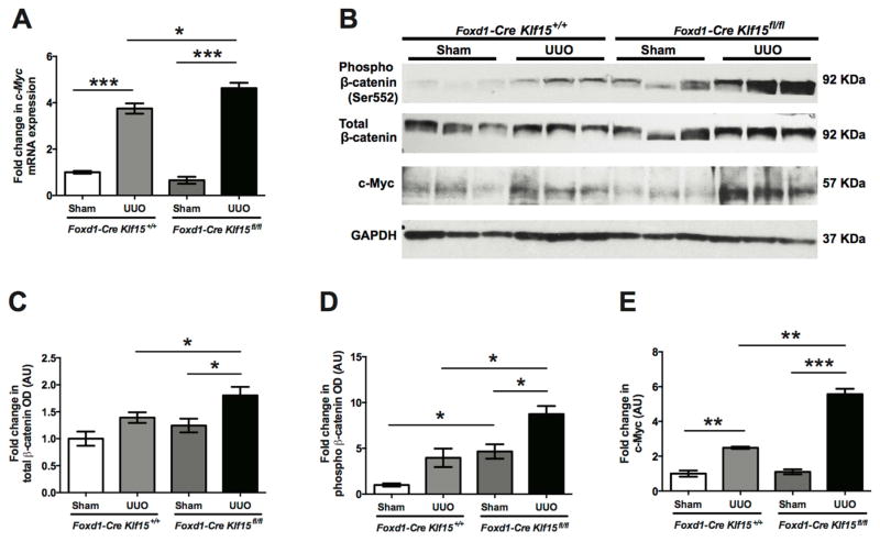
Figure 4. Activation of Wnt/β-catenin signaling in UUO-treated Foxd1-Cre Klf15fl/fl mice.
(A) RNA was extracted from total kidney cortex and RT-PCR was performed for c-Myc expression from 12-week-old Foxd1-Cre Klf15fl/fl and Foxd1-Cre Klf15+/+ mice treated with sham or UUO for 3 days. (n=6, *p<0.05, ***p<0.001, Kruskal-Wallis test with Dunn’s post-test). (B–E) Western blot was also performed on total kidney cortex for phospho-β-catenin (Ser552), total-β-catenin, c-Myc, and GAPDH. Representative blots from three independent experiments are shown. Densitometry analysis was performed to quantify protein expression. (n=3, *p<0.05, **p<0.01, ***p<0.001, Kruskal-Wallis test with Dunn’s post-test).

