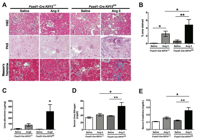
Figure 6. Loss of Klf15 in Foxd1+ stromal cells exacerbates AngII-induced fibrosis.
Age-matched 12-week-old Foxd1-Cre Klf15fl/fl and Foxd1-Cre Klf15+/+ mice were concurrently treated with saline or AngII continuous infusion for 6 weeks. (A) All mice were sacrificed and renal cortex fixed for histology. Hematoxylin and Eosin (H&E), Periodic acid-Schiff (PAS), and Masson’s Trichrome staining was performed to evaluate for tubulointerstitial changes. The representative images from 6 mice in each group are shown (X 20). (B) % area fibrosis from Masson’s Trichrome stain is shown. (C) Urine albumin to creatinine (cr), (D) Serum urea nitrogen, and (E) Serum creatinine were measured in all 4 groups 6 weeks post treatment. (n=6, *p<0.05, **p<0.01, Kruskal-Wallis test with Dunn’s post-test).

