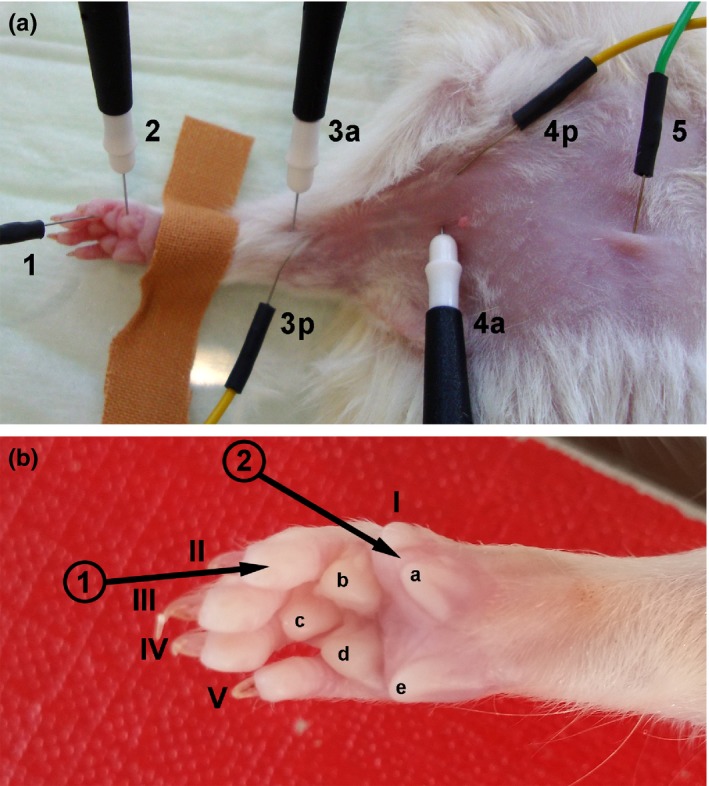
Figure 2.

Photographs demonstrating the ventral view of electrode positioning during noninvasive electrophysiological recordings and of the magnified palmar view of the right paw to clarify exact positioning of the recording electrode. (a) Proximal stimulation was achieved by introducing active (4a) and passive (4p) stimulation electrodes close together in the axillary region while distal stimulation was performed by placing active (3a) and passive (3p) stimulation electrodes close together at the elbow. The ground electrode (5) was subcutaneously introduced above the sternum. (b) As indicated by the black arrows, the recording electrode (2) was positioned at the thenar muscle between the rudimentary thumb (I) and the medial walking pad (a), while the reference electrode (1) was placed in the finger tip of the second digit (II)
