Fig. 1.

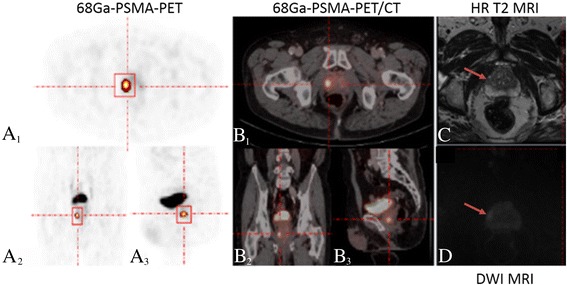
Example of tumor heterogeneity for the asphericity assessment in a patient diagnosed with a Gleason score 6 prostate cancer. A–D Example of a patient diagnosed with a Gleason score 6 prostate cancer in the right peripheral zone of the apex of the prostate. A, B Corresponding orientations (transversal (A 1), coronal (A 2), and sagittal (A 3)) are shown for 68Gallium-labeled prostate-specific membrane antigen positron emission tomography, fused with computed tomography (B 1–3). Based on positron emission tomography, an avid tumor was delineated in all three planes using the rover software. C, D Corresponding magnetic resonance imaging in the high-resolution T2 turbo spin echo (C) and diffusion-weighted images (D) in axial images confirm the presence of the malignant lesion at the respective location. HR: high-resolution, DWI: diffusion-weighted imaging
