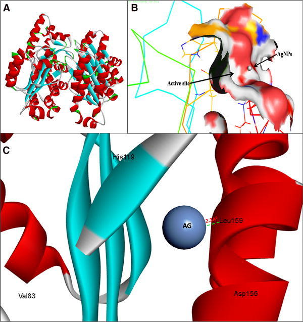
Fig. 2.

3D structure of transcriptional activator protein LasR (a); binding of AgNPs with the active site of LasR (b); close view of catalytic site of LasR, with bound Ag represented as blue sphere. Amino acids residues (Leu159) of LasR involved in the interaction with AgNPs (c). LasR active site has been depicted by PyMol viewer. Interaction analysis of AgNPs with LasR has been explored through PatchDock
