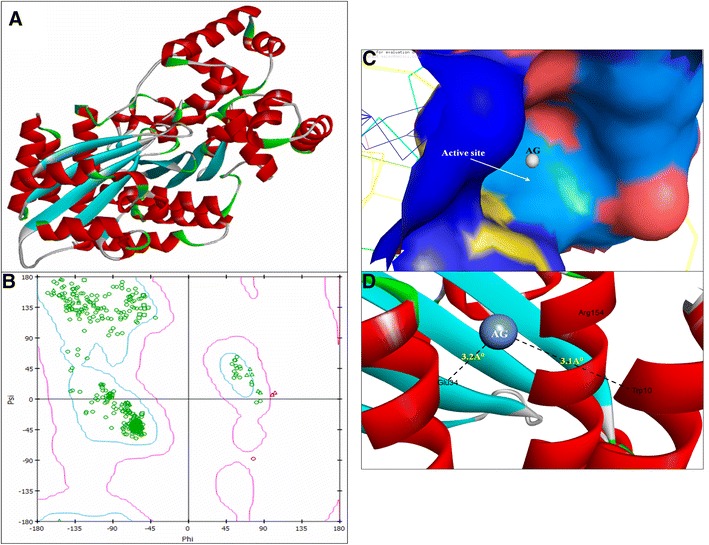
Fig. 4.

3D structure of regulatory protein RhlR (a); RAMPAGE validation of conformation of RhlR (b); binding of AgNPs with the active site of RhlR (c); close view of catalytic site of RhlR, with bound Ag represented as blue sphere. Specific residues (i.e., Trp10 & Glu34) of RhlR involved in the interaction with AgNPs (d). RhlR active site has been depicted by PyMol and the interaction analysis of AgNPs with RhlR has been explored by PatchDock
