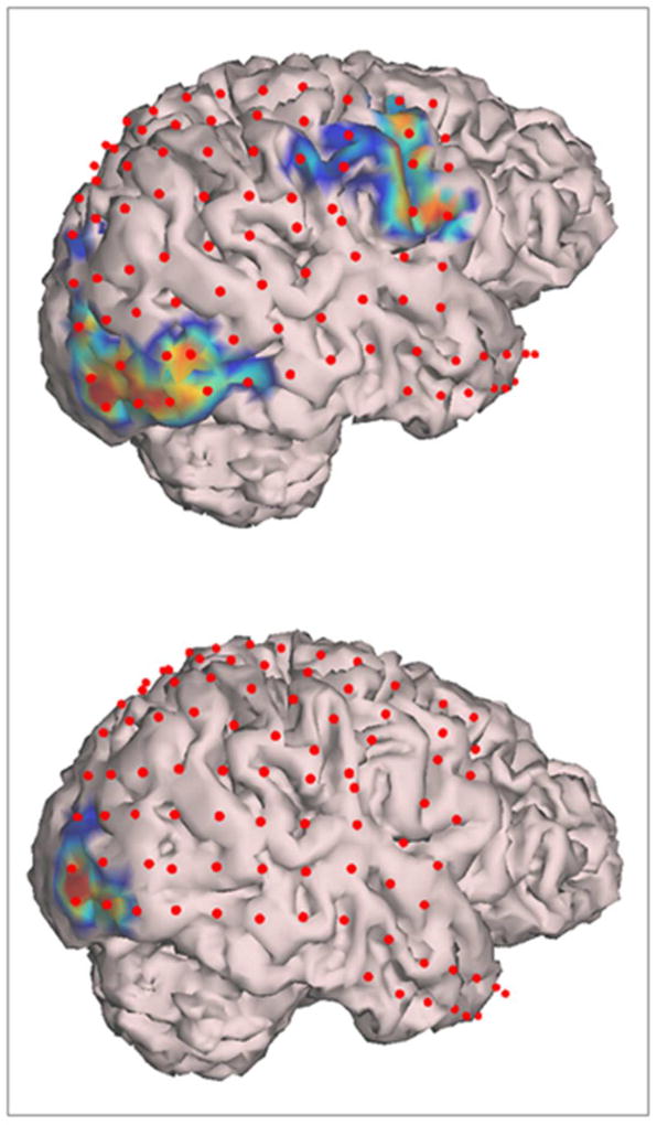
Figure 2.
Segmented cortical model derived from patient R3’s brain MRI with superimposed subdural electrodes (red dots), and high-γ modulations (HGM) for overt (top panel) and covert (bottom panel) visual naming. Note the activation of oral/facial motor cortex with overt (top panel) but not covert (top panel) naming. Also note the activation of visual cortex in occipital lobe in both conditions.

