
FIG 2.
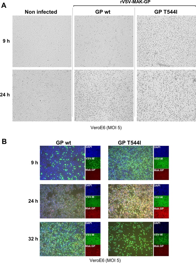
Phenotypic differences between rVSV-MAK-GP and mutant rVSV-MAK-GP T544I viruses. (A) Cells infected with mutant rVSV-MAK-GP show more rapid induction of CPE than cells infected by the wild type. Vero E6 cells were infected at a multiplicity of infection (MOI) of 5 and imaged by bright-field microscopy at the indicated times. Images are representative of the monolayer. (B) Immunofluorescence staining showing more rapid viral protein expression in cells infected with the mutant rVSV-MAK-GP T544I virus. Vero E6 cells infected at an MOI of 5 were stained at the indicated times for VSV M protein, Makona GP1,2 protein, and nuclei (4′,6′-diamidino-2-phenylindole [DAPI]). The large boxes show merged images, and the smaller boxes show separate channels. Images were captured with a 10× objective under identical exposure settings.