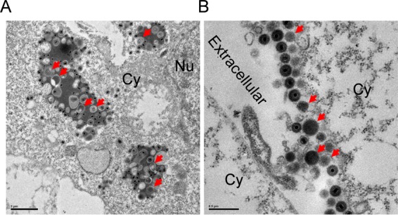
FIG 2.

VLVs are generated in multivesicular bodies. (A) Formation of VLVs in cells lytically infected with MHV-68. NIH 3T3 cells were first infected with MHV-68 and then examined by plastic embedding, ultrathin sectioning, and transmission electron microscopy. Red arrows indicate VLVs. Cy, cytoplasm; Nu, nucleus. (B) Release of VLVs into extracellular space.
