Fig. S1.
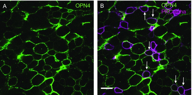
Subretinal vector delivery leads to appropriate membrane localization of human melanopsin and bipolar cell transduction in end-stage retinal degeneration. Staining of a retinal flatmount with OPN4 antibody (green) illustrates its location at the edges of INL cells, consistent with membrane localization of human melanopsin (A). Colabeling with PKCα antibody (purple) demonstrates successful transduction of a number of rod bipolar cells (arrows, B) following subretinal delivery of melanopsin vector. (Scale bar, 10 μm.)

