Fig. S2.
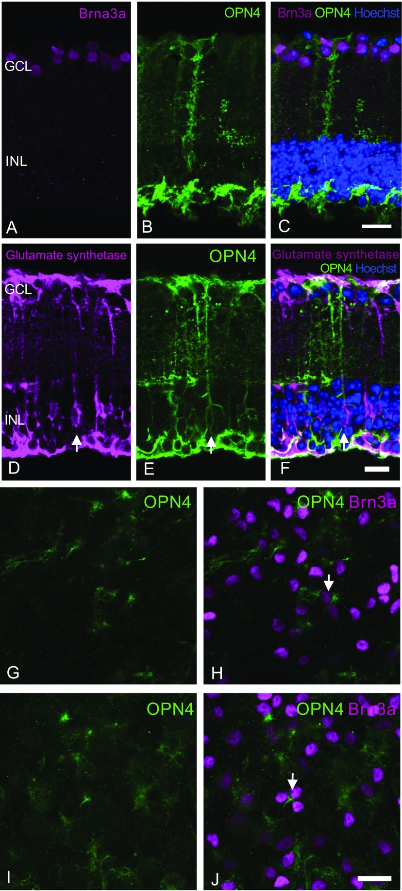
No significant transduction of cell bodies within the ganglion cell layer is seen following vector delivery. Retinal sections were stained with Brn3a (purple, A) and OPN4 (green, B) antibodies and overlaid with Hoechst nuclear stain (blue) to identify any ganglion cell transduction by the OPN4 vector (C). Examination of histological sections showed melanopsin staining within the ganglion cell layer, but this appeared to be due to labeling of Müller cell end feet [stained using glutamate synthetase antibody, purple, overlaid with Hoechst nuclear stain (blue), D–F]. Staining of retinal flatmounts (G–J) with Brn3a (purple) and OPN4 (green) antibodies did not show significant human melanopsin staining of cell bodies within the ganglion cell layer, suggesting that ganglion cells were not transduced by subretinal delivery of the OPN4 vector. Some human melanopsin-expressing axonal processes did appear to meet Brn3a-positive ganglion cells (arrows). These could be human melanopsin-expressing bipolar cells synapsing with ganglion cells, or Müller cell end feet surrounding them. (Scale bar, 25 μm.)

