Fig. S4.
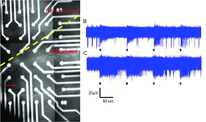
MEA recordings from treated retinas demonstrate a range of response kinetics. Four months after subretinal injection of OPN4 vector, multielectrode array (MEA) recordings were performed on ex vivo retinas of treated eyes and age-matched untreated controls. Following completion of recordings, DsRed fluorescence was visualized in treated eyes with the retina in situ over electrodes. This allowed areas of retina transduced by vector to be identified (A, the image shown is a composite illustrating the area covered by one treated retina overlying the MEA electrodes; arrow indicates dots that are DsRed-expressing cells). To examine duration of response, the time to half decay [time from maximal firing rate to half-response (t1/2)] of light responses recorded from treated retina following a 60-s light pulse of 1.3 × 10¹⁵ photons⋅cm−2⋅s−1 intensity was examined. This ranged from 6 to 147 s (mean, 51.1 ± 5.5 s; n = 54) and was not significantly different from values obtained from untreated controls where responses ranged from 14 to 144 s (mean, 64.8 ± 8.2 s; n = 25; P = 0.165, two-tailed unpaired t test). Time to half-decay was shorter following light pulses of 2-s duration, typically lasting 1–24 s in treated retina (mean, 6.9 ± 1.3 s; n = 16) and 1–12 s in untreated retina (mean, 6.4 ± 1.9 s; n = 5; P = 0.84, two-tailed unpaired t test). Responses with transient (B) and sustained kinetics (C) were also seen following a 2-s light stimulus. Examples of raw data obtained from individual electrodes recorded from a treated retina are shown following 480-nm light pulses of 2-s duration that were repeated every 60 s.

