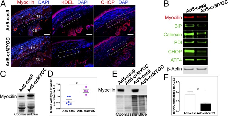
Fig. 3.
CRISPR efficiency in vivo in mice and ex vivo in human eyes. (A) Representative image showing that Ad5-crMYOC treatment reduces myocilin (red), KDEL (red), and CHOP (red) levels in the TM (rectangular box) of Tg-MYOCY437H mice (n = 3). C, cornea; CB, ciliary body. (Scale bar: 50 µm.) (B) Representative image of WB samples from a TM ring showing that Ad5-crMYOC treatment decreases myocilin levels and associated BiP, Calnexin, PDI, ATF4, and CHOP protein levels (n = 9). (C) Representative image showing increased secretion of mouse WT myocilin into the aqueous humor of Ad5-crMYOC–treated eyes compared with Ad5-cas9–treated eyes. (D) Densitometry showing a significant increase in the levels of secreted mouse WT myocilin in Ad5-crMYOC–treated eyes of Tg-MYOCY437H mice (n = 4). (E) inhibition of myocilin secretion by Ad5-crMYOC–mediated myocilin knockdown in a human ex vivo anterior segment perfusion culture (n = 2). Coomassie blue staining was performed to ensure relatively equal loading. (F) Decreased myocilin (MYOC) mRNA expression in human ex vivo anterior segment perfusion culture by Ad5-crMYOC treatment. Error bars represent SEM. *P < 0.05.

