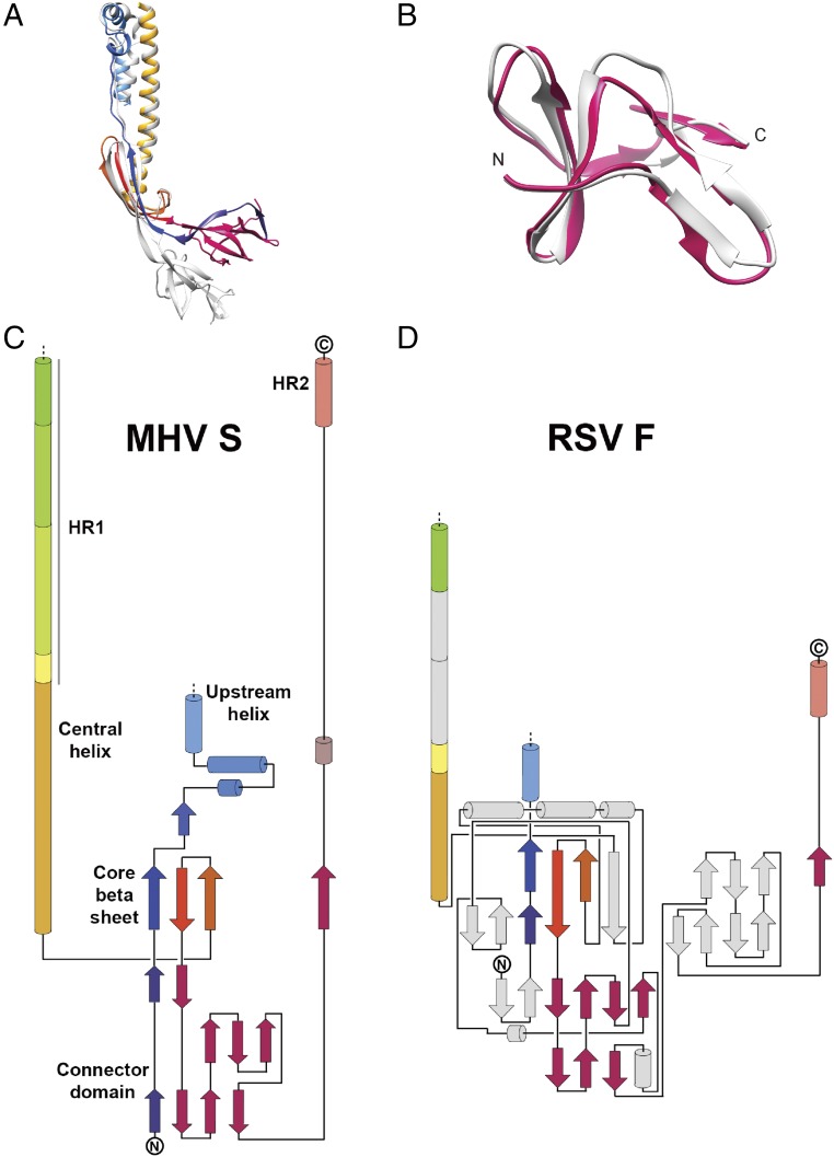
Fig. S4.
Comparison of the MHV S postfusion S2 structure with other fusion proteins. (A) Superimposition of the HCoV-NL63 S2 (gray) and MHV postfusion S2 (colored as in Fig. 2) subunits. Only the upstream and central helices, core β-sheet, and connector domain are shown. (B) Superimposition of the HCoV-NL63 S2 (gray) and MHV postfusion S2 (pink) connector domains emphasizing their structural conservation. (C and D) Topology diagrams comparing postfusion MHV S2 and RSV F glycoproteins. Conserved elements in the RSV F subunit are shown using the same colors; nonconserved elements are shown in gray.

