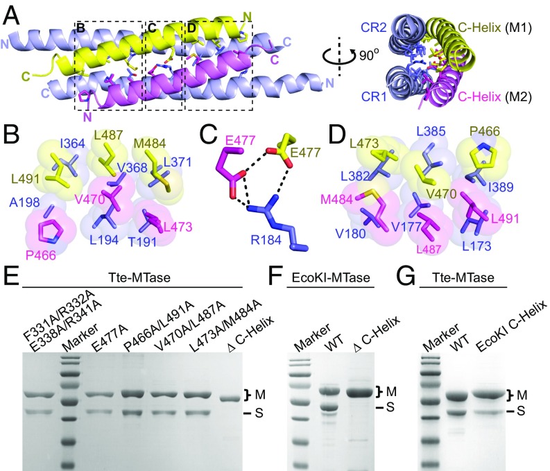
Fig. 2.
Four-helix bundle interface between S and M subunits. (A) Ribbon diagram of the four-helix bundle structure consisting of the CR regions (light blue) from the S subunit and two C-helices (yellow and violet) from the M subunits. The key residues are shown in stick representation. (B–D) Detailed interactions within the four-helix bundle interface as highlighted by the dashed boxes in A. The hydrophobic interactions are shown in space-filling presentation (B and D). The hydrogen bonds are shown as thick black dashed lines (C). Same color code as in A. (E–G) SDS/PAGE results of the coexpression of the wild-type S subunit (no tag) and different M subunits (His tagged) as indicated for both Tte (E and G) and EcoKI (F) systems. The SDS/PAGE gels were stained with Coomassie blue.

