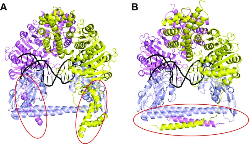
Fig. S5.
(A) The negative-stain EM closed model of the EcoKI-MTase. Same color code for the proteins as in Fig. 1. The DNA is shown in black ribbon. The red circles indicate the C-helix regions in the wrong conformation. (B) The updated closed model of the Tte-MTase. The S and M subunits of the EcoKI enzyme in A were replaced by their Tte counterparts by superimposition. The red circle indicates the revised four-helix bundle region based on the crystal structure.

