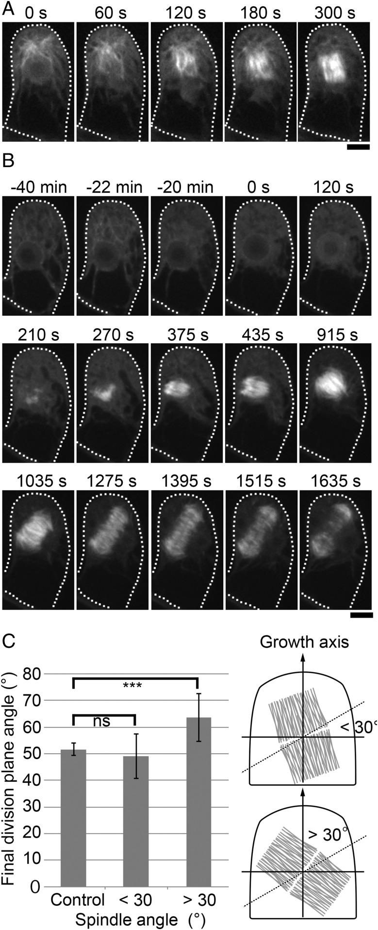
Fig. 2.
The gametosome plays a critical role in spindle orientation. (A) Time-lapse imaging of mCherry–α-tubulin during the first division of the gametophore initial. Images were recorded every 15 s. Time 0 corresponds to the timing of NEBD. MTs emanated from the gametosome penetrate into the nucleus upon NEBD. Subsequent MT amplification and bipolarization leads to metaphase spindle formation (300 s). (B) Time-lapse imaging of mCherry–α-tubulin in a cell transiently treated with oryzalin. Before the first division in the gametophore initial, cells were treated with 20 µM oryzalin (−22 min), which destabilized the gametosome. Oryzalin was washed out upon NEBD (time 0), and images were recorded every 15 s. MTs were nucleated in a gametosome-independent manner and a bipolar spindle was formed (375 s). However, the orientation of the spindle was ectopic, as was also the angle of the expanding phragmoplast. Cells are outlined with dotted lines. (Scale bars, 10 µm.) (C) Measurement of final division plane angle in cells transiently treated with oryzalin. The cells were classified according to their spindle angles, which were within 30° (n = 5) or over 30° (n = 5), after oryzalin washout. The final division plane angles of classified cells were compared with control cells (n = 5) using t test. ***P < 0.02; ns, P > 0.5. Illustration of the spindle angle (within 30° or over 30°) on the right of graph. The dotted line shows 30°, and the gray structure indicates the spindle.

