Fig. 3.
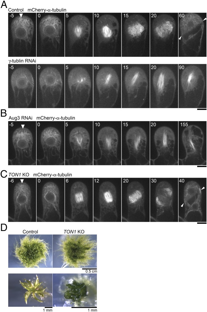
γ-Tubulin is required for gametosome formation. (A and B) Time-lapse imaging of mCherry–α-tubulin during the first division of the gametophore initial in control, γ-tubulin RNAi, and Aug3 RNAi lines. Images were recorded every 5 min. Time 0 corresponds to the timing of NEBD. Arrowheads indicate gametosomes. (Scale bars, 10 µm.) (C) Time-lapse imaging of mCherry–α-tubulin during the first division in the gametophore initial of the PpTON1-deletion line. Images were recorded every 2 min. Time 0 corresponds to the timing of NEBD. The gametosome structure appears normally in prophase (arrowhead). (Scale bar, 10 µm.) (D) Abnormal gametophore morphology resulting from the PpTON1 deletion, consistent with previously reported data (30). Arrowheads in the final frames of A and C show the final division plane orientation.

