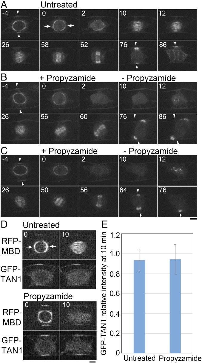
Fig. 6.
Polar cap-dependent spindle orientation contributes to division plane control in tobacco BY-2 cells. (A–C) Time-lapse imaging of YFP-β-tubulin in a control untreated cell (A) and upon transient treatment with propyzamide (B and C). Before NEBD (time 0), cells were treated with 10 µM propyzamide that destabilized the polar caps. Propyzamide was washed out after cells were allowed to progress past NEBD for 10 min, and images were recorded every 30 s. MTs were nucleated in a polar cap-independent manner and a bipolar spindle formed (26 min). However, orientation of the spindle was random. In the cell presented in C, the division plane was eventually corrected by phragmoplast guidance toward the region where PPB was formerly present (50–64 min), whereas in B the guidance mechanism was not sufficient to correct the plane (76 min). Arrowheads indicate the PPB and the corresponding region marked by PPB before NEBD. Arrows indicate polar caps. (Scale bar, 10 µm.) (D) Representative images of BY-2 cells expressing RFP-MBD and the CDZ marker GFP-TAN1 in a control untreated cell and upon transient treatment with propyzamide. Time is indicated in minutes. (Scale bar, 10 µm.) (E) Quantification of GFP-TAN1 intensity at the CDZ of untreated and treated BY-2 cells. The ratio of the fluorescence intensity between time point 0 min and time point 10 min is shown with SD (n = 10). Arrows indicate the polar caps.

