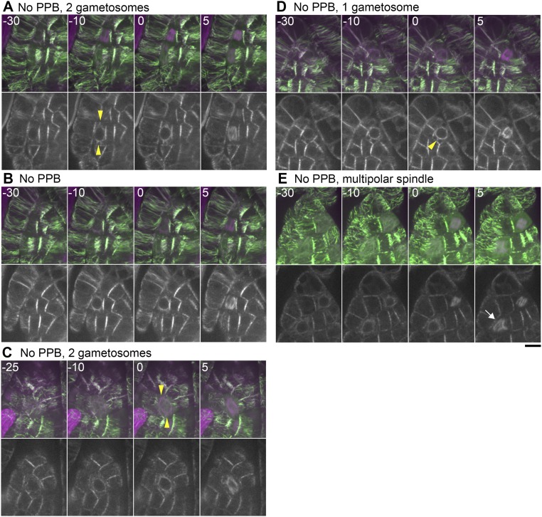
Fig. S5.
Gametosome formation during cell division in leaf-like gametophore cells without clear PPBs. (A–E) Time-lapse imaging of Citrine-MAP65d (green) and mCherry–α-tubulin (magenta) in mature gametophore cells. Images were acquired every 5 min with 16 z-sections (separated by 1 µm) and displayed after maximum projection (Top, merged) or as single median slices (Bottom, grayscale). Time 0 corresponds to the timing of NEBD. White and yellow arrowheads indicate the PPB and gametosome structures, respectively. The arrow indicates a multipolar spindle. (Scale bar, 10 µm.)

