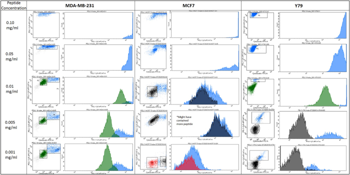
Figure 3.
Titrations of RK-10-Cy5 in Cell Lines using flow cytometry. Cell lines expressing high PD-L1 (MDA-MB-231) and low PD-L1 (MCF7, Y79) were incubated with fluorescent RKC-10-Cy5 peptide and anti-cytokeratin for 1 hour at shown concentrations, then analyzed using flow cytometry. In some cases we can distinguish populations of PD-L1 expression, separated by color. Higher concentrations of peptide were unable to distinguish expression, while concentrations of 0.005 and 0.001 mg/mL peptide were selected as ideal for flow cytometry.

