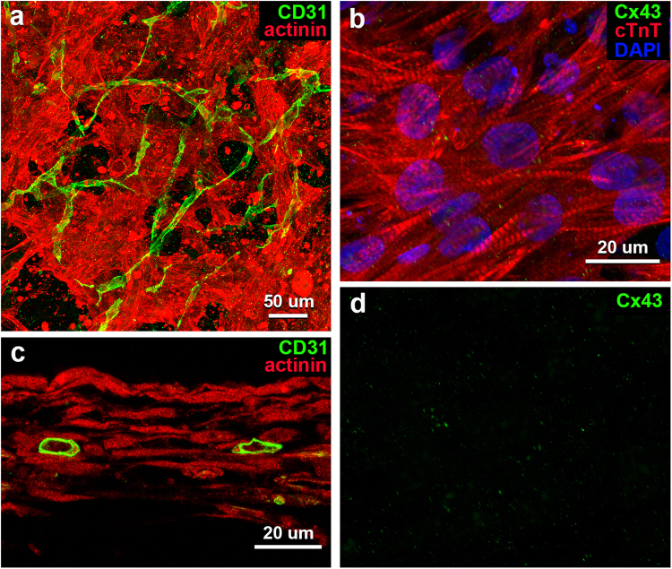
Figure 1.
Light microscopic images of whole-mount preparations (a,b) and a tissue section (c) of vascularized iPSC-CM 3D-tissues 4 days after construction. (a,c) Immunostaining for sarcomeric α-actinin (red) and CD31 (green) shows a meshwork of iPSC-CMs preparing myofibrils and a network of endothelial tubes with lumens. (b) Immunostaining for cTnT (red) and Cx43 (green) shows sarcomeres in iPSC-CMs, and a small amount of reaction products for Cx43 on the cells. Cellular nuclei are stained with DAPI. (d) An image of reaction products for Cx43 distilled from b.

