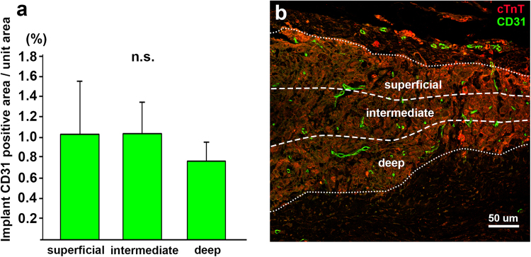
Figure 4.
Comparison of vascular distribution in iPSC-CM 3D-tissues 28 days after implantation to rat infarcted heart. (a) The ratio of the CD31-immunopositive vascular area to unit area in the deep region (0.76 ± 0.19%; n = 3, total 6 fields) is lower than that in the intermediate (1.03 ± 0.31%; n = 3, total 6 fields) or superficial (1.04 ± 0.52%; n = 3, total 6 fields) region within the implant, though there is no statistical difference among each region. (b) A representative image of three divided regions in the implant and vascular distribution in each region. Immunostaining for cTnT (red) and multi-species CD31 (green) on a transverse section of the iPSC-CM 3D-tissue implanted to the infarcted rat heart. The pericardium is located at the top of the image.

