Fig. 3.
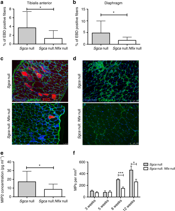
Pathological parameters are rescued in dystrophic mice lacking Nfix. a Percentage of EBD positive myofibers in tibialis anterior muscles at 8 weeks; N = 19 Sgca null and 9 Sgca null:Nfix null mice; mean ± SD; t test, *P < 0.05. b Percentage of EBD positive myofibers in diaphragms at 8 weeks; N = 19 Sgca null and 11 Sgca null:Nfix null mice; mean ± SD; t test, *P < 0.05. c Immunofluorescence for laminin (green) and EBD (red) on diaphragms at 8 weeks. Hoechst (blue) stains nuclei. Scale bar 50 μm. d Immunofluorescence showing collagen I (green) deposits in tibialis anterior sections. Scale bar 100 μm. e MIP2 ELISA assay on gastrocnemius muscles at 8 weeks; N = 18 Sgca null and 7 Sgca null:Nfix null mice; mean ± SD; t test, *P < 0.05. f Quantification of the immunofluorescence staining for F4/80, marker of macrophages (MPs), on tibialis anterior muscle sections at different time points. N = 5 Sgca null and 5 Sgca null:Nfix null at 3 weeks, N = 3 Sgca null and 3 Sgca null:Nfix null at 5 weeks, N = 8 Sgca null and 8 Sgca null:Nfix null at 8 weeks, and N = 12 Sgca null and 7 Sgca null:Nfix null at 12 weeks. Mean ± SEM; t test, *P < 0.05, ***P < 0.001

