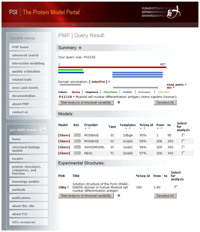
Fig. 5.
Protein Model Portal (PMP) query results for the human myeloid cell nuclear differentiation antigen protein (UniProt P41218 (94, 95), represented as a red bar). For the first 90 residues of this protein an experimentally solved structure (green bar) is deposited in the PDB database (PDB ID 2dbg (102)). The protein structure corresponds to the PPAD_DAPIN N-terminal domain of the protein. For the C-terminal HIN domain, three homology models are obtainable from the PMP model providers ModBase, SWISS-MODEL and NESG. Below the graphical representation a list of models and information about the structure is available. Additional information is accessible by clicking the corresponding model or PDB ID links. A subset of models or structures can be selected for further structural comparison.

