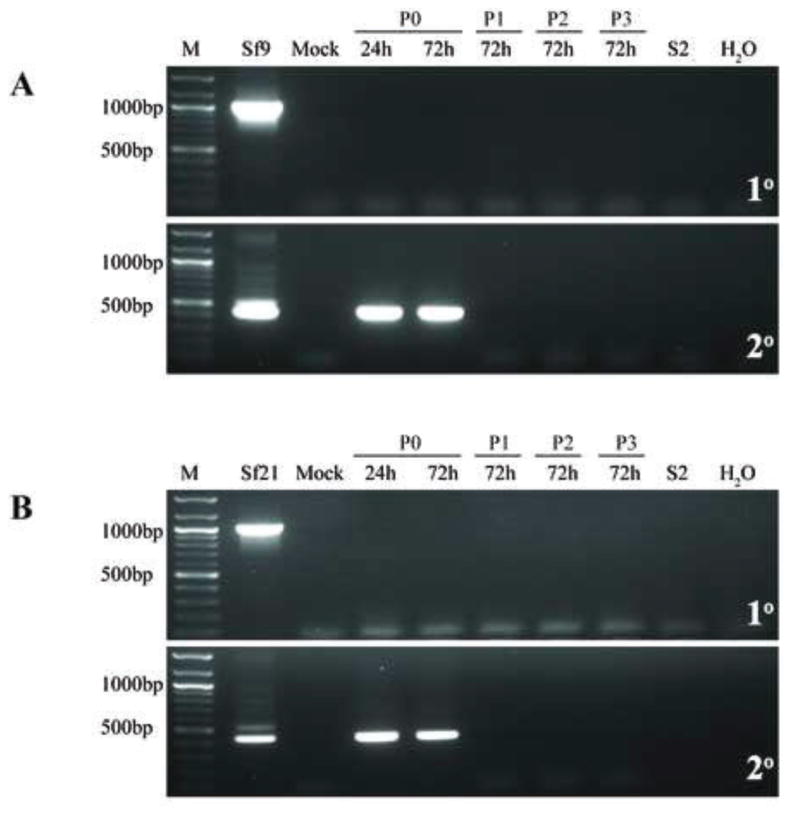
Fig. 7.
Infectivity of representative Sf-rhabdovirus variants in MDBK cells. MDBK cells were inoculated with fresh C-TNMFH (Mock) or C-TNMFH media filtrates from (A) Sf9 or (B) Sf21 cells and total RNAs were isolated from cell extracts at various times and passages after inoculation and assayed by primary RT-PCR (upper panels) and RT-PCR/nested PCR (lower panels) using N-specific primers, as described in the legend to Fig. 5. Positive and negative controls for the RT-PCRs were also performed as described in the legend to Fig. 5 and the lane marked M shows the 100-bp markers, with selected sizes indicated on the left.

