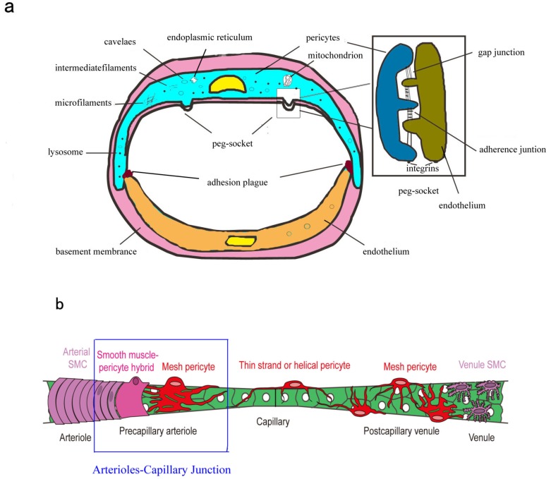
Fig. (2).
Brain pericytes anatomy. (a) Ultrastructural of brain pericytes. Brain pericytes nucleus is relatively large, kidney-shaped and protrudes into tubal antrum. The small amounts of perinuclear cytoplasm usually contain mitochondria, endoplasmic reticulum and lysosomes. Contractile microfilaments (containing actin, myosin and tropomyosin) and intermediate filaments are also observed in these cells. Numerous caveolae principally locate in abluminal pericytes surface and single caveolae usually appear in the processes of pericytes. In the areas lacking BM, pericytes and ECs form direct cell-to-cell contacts with each other by interdigitation, referred to as “peg-and-socket” contacts. These contacts include the connexin-43 hemichannels (CX-43) mediated gap junctions and N-cadherin based adherence junctions. The other type of contacts is “adhesion plaques”. (b) Topology and morphology of brain pericytes. Schematic showing the continuum of mural cell types along the cerebral microvessels. Brain arterioles wrapped by a continuous of vascular smooth muscle cells (vSMCs) further ramify and transit into smaller precapillary arterioles. The mural cells located in the point of transition show a mixed phenotype of vSMCs and pericytes, termed as “smooth muscle-pericytes hybrids”. Another type of pericytes at the precapillary arterioles, referred to as “mesh pericytes”, exhibit more interlocking mesh progresses. Pericytes in capillary beds have protruding cell bodies with thin strand or helical primary processes that run parallel to the long axis of mid-capillary tube. Mesh pericytes with many slender, branching processes become more prevalent on postcapillary venules. The boexs represent “arterioles-capillary junction”. (Reproduced from Hartmann et al. [32] with permission).

