Figure 2.

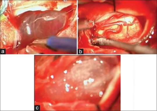
(a) Intraoperative view showed subdural thick scars and arachnoid cyst. (b) The spinal cord is reconstructed after the dissection. (c) The platelet-rich fibrin + stem cells in place at the end of surgery

(a) Intraoperative view showed subdural thick scars and arachnoid cyst. (b) The spinal cord is reconstructed after the dissection. (c) The platelet-rich fibrin + stem cells in place at the end of surgery