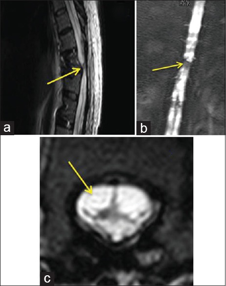
Figure 5.

(a) Magnetic resonance imaging - sagittal cuts of the dorsal spine of case 2 showed transection of the spinal cord at the level of D10–D11. (b) Magnetic resonance imaging myelogram showed spinal cord transaction at the level of D10–D11. (c) Magnetic resonance imaging - axial cuts of D10 demonstrated severely damaged and transected spinal cord
